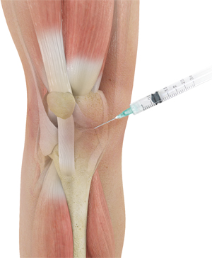
Ultrasound can be used to help physicians to perform image guided injections. It means that the treating physician would use ultrasound to look at the anatomy of the area to be injected and use the guidance of image for performing the injection more precisely.
Benefits: Performing more precise injections by seeing where the needle is going to. Observing real time injection procedure while it happens to now when to start and when to end. To make sure the needle is not in blood vessel. To differentiate different types of tissues under the ultrasound image. To be able to diagnose some issues with tissues before starting treatments. It helps to get to the exact desired point of injection.
Limitations: The deeper the lesions, the lower the image resolution under ultrasound would be. The ultrasound’s benefits are dependent to the operator’s techniques. Physician needs to know about the local anatomy and understand the sonoanatomy. Ultrasound cant penetrate the bones and creates shadows when it reaches the bones. If bones are to be evaluated, X-Ray needs to be used.
In summary, it is a good tool in hand for when anatomical landmarks alone cannot be trusted for precision or safety of an injection.
Ultrasound Guided Injections


